What Should You Know Before Going for Low Exposure Digital Radiographs?

When it comes to dental health, advancements in technology have revolutionized the way we diagnose and treat oral issues. One such advancement is Low Exposure Digital Radiographs (LEDR), a cutting-edge method that offers numerous benefits over traditional X-rays. As dental professionals at Lake Success Dental Group, located at 3333 New Hyde Park Road, Suite G5 New Hyde Park, NY 11042, we prioritize patient safety and health, making LEDR an essential part of our practice.
In this blog, we’ll delve into everything you need to know about Low Exposure Digital Radiographs, including their benefits, how they work, the technology behind them, and practical tips for patients.
What Are Low Exposure Digital Radiographs?
Low Exposure Digital Radiographs are advanced imaging techniques that use digital sensors instead of traditional photographic film to capture detailed images of your teeth and jaw. These radiographs require significantly less radiation than conventional X-rays, making them a safer option for patients.
Benefits of Low Exposure Digital Radiographs
- Reduced Radiation Exposure: LEDR uses up to 90% less radiation compared to traditional X-rays. This is especially beneficial for patients who require frequent imaging.
- Enhanced Image Quality: The digital sensors in LEDR provide high-resolution images. We can easily enhanced and enlarged for better diagnosis.
- Immediate Results: Digital radiographs are instantly available for viewing, allowing for quicker diagnosis and treatment planning.
- Environmentally Friendly: Unlike traditional X-ray films, LEDR does not require chemical processing, making it a more eco-friendly option.
- Improved Storage and Sharing: Digital images can be easily stored in patient records and shared with other dental specialists if needed.
How Do Low Exposure Digital Radiographs Work?
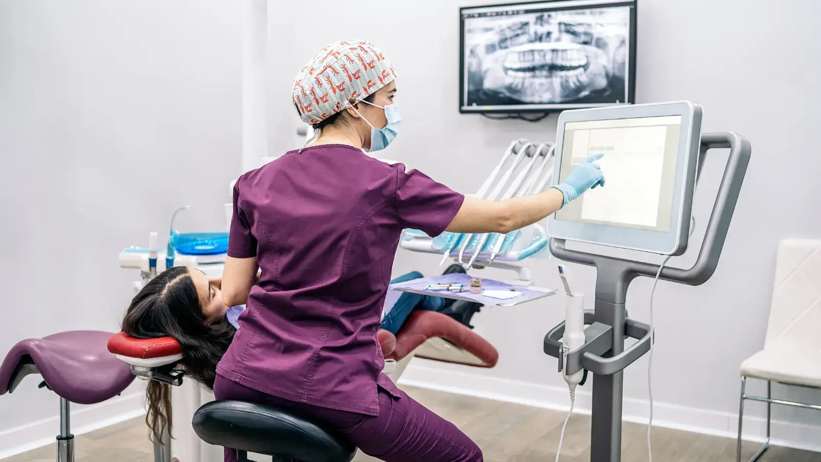
LEDR technology involves the use of a digital sensor placed inside the mouth, which captures the image when exposed to a small amount of X-ray radiation. The sensor is connected to a computer that processes the image almost instantaneously, allowing the dentist to view the results on a monitor.
The Technology Behind LEDR
The key component of LEDR is the digital sensor. There are two main types of sensors used:
- Charge-Coupled Device (CCD): This type of sensor captures X-ray images with high sensitivity and resolution.
- Complementary Metal-Oxide-Semiconductor (CMOS): CMOS sensors are known for their efficiency and lower power consumption while providing excellent image quality.
Both types of sensors convert the X-ray photons into electrical signals, which are then processed by the computer to produce detailed digital images.
Research Data and Facts
Numerous studies have highlighted the benefits of LEDR in dental practices. Here are some key findings:
- A study published in the Journal of Dentistry found that LEDR reduces radiation exposure by 70-90% compared to conventional X-rays.
- According to the American Dental Association (ADA), digital radiographs have a higher diagnostic accuracy, especially for detecting dental caries and periodontal diseases.
- Research from Harvard School of Dental Medicine indicates that the enhanced image quality of LEDR can lead to more accurate diagnoses and better treatment outcomes.
Tips and Tricks for Patients
Preparing for Your LEDR Appointment
- Inform Your Dentist: Always inform your dentist about any existing medical conditions or if you are pregnant.
- Stay Still During the Procedure: To ensure clear images, try to remain as still as possible while the sensor is in your mouth.
- Follow Instructions: Your dental professional will guide you on how to position your head and bite down on the sensor.
After the Procedure
- Review the Images with Your Dentist: Take the time to review the digital images with your dentist. This is a great opportunity to ask questions and understand your dental health better.
- Follow-Up Care: Based on the findings from the radiographs, follow any recommended treatment plans or preventive care routines.
FAQs About Low Exposure Digital Radiographs
Are Low Exposure Digital Radiographs Safe?
Yes, LEDR are safe and significantly reduce radiation exposure compared to traditional X-rays. The technology adheres to strict safety standards to ensure patient well-being.
How Often Should I Get Digital Radiographs?
The frequency of digital radiographs depends on your dental health needs. Typically, a full set of radiographs is recommended every 1-2 years, with bitewing radiographs taken annually for patients with good oral health.
Do Digital Radiographs Hurt?
The procedure is generally painless. Some patients may experience slight discomfort from the sensor placed in the mouth, but it is brief and minor.
Can Children Get Low Exposure Digital Radiographs?
Absolutely! LEDR is safe for children and is often used to monitor the development of their teeth and detect any early signs of dental issues.
The Future of Dental Imaging
As technology continues to advance, the future of dental imaging looks promising. Innovations such as 3D digital radiography and AI-enhanced diagnostics are on the horizon. Which are promising even more precise and efficient dental care.
Conclusion
Low Exposure Digital Radiographs are a remarkable advancement in dental imaging, offering numerous benefits over traditional X-rays. They provide high-quality images with minimal radiation exposure, ensuring a safer and more efficient diagnostic process. By understanding how LEDR works and following the tips provided, patients can make informed decisions about their dental health.
We, at Lake Success Dental Group, are dedicated to using the best technology to provide top-notch dental care for our patients. If you have any questions or need to schedule an appointment, don’t hesitate to contact us at 3333 New Hyde Park Road, Suite G5 New Hyde Park, NY 11042. Your dental health is our priority, and we look forward to helping you achieve a healthy, beautiful smile.